Facts about cardiovascular disease
Many patients do not fully understand the risks associated with cardiovascular disorders or which factors contribute to the disease.
Age and family genetics can contribute to the likelihood of developing arterial problems but there are also contributing factors that patients can control:
Smoking
Lack of exercise
Obesity
Excessive stress
High-fat diet
Once diagnosed, arterial problems may be treated via a plan that a physician will develop. The plan may include:
An exercise routine to improve circulation and lower high blood pressure.
Medication, such as blood thinners.
Surgical procedures such as bypass, AAA graft, angioplasty, stents, or endarterectomy.
Below you will find an explanation of three common arterial problems; carotid artery blockage, abdominal aortic aneurism (AAA), and peripheral artery disease (PAD).
Carotid Artery Blockage
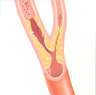
Excessive plaque buildup in carotid arteries can
restrict free flow of blood to the brain. It may also cause blood clots to
form which can travel in the blood to the brain and may block or restrict blood
supply to smaller vessels. This condition may result in a stroke.
Treatment of this condition depends on the amount
of plaque buildup and the patient's symptoms. A physician may order a
carotid duplex ultrasound to determine the severity of plaque buildup.
Treatment may range from making lifestyle changes to medication or surgical
procedures.
A carotid artery duplex exam shows the amount (if
any) of plaque buildup in the carotid arteries, the type of plaque, and the
velocity of blood flow through the arteries.
Abdominal Aortic Aneurism (AAA)
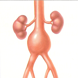 The
abdominal aorta carries blood from your heart, through your abdomen, to the
lower body. A life-threatening condition may occur when the wall of the
aorta is weakened and begins to stretch as shown in the picture to the left.
If the artery is stretched too much, the aorta may burst.
The
abdominal aorta carries blood from your heart, through your abdomen, to the
lower body. A life-threatening condition may occur when the wall of the
aorta is weakened and begins to stretch as shown in the picture to the left.
If the artery is stretched too much, the aorta may burst.
Unfortunately, there often aren't any symptoms.
AAA is usually found when a patient undergoes an unrelated test. Once a
patient is diagnosed with AAA, a physician will order an aorta duplex ultrasound
to measure the size of the aneurism.
Treatment depends on the size of the aneurism and
may range from monitoring (in case of a small aneurism) to endovascular surgical
procedures.
Peripheral Artery Disease (PAD)
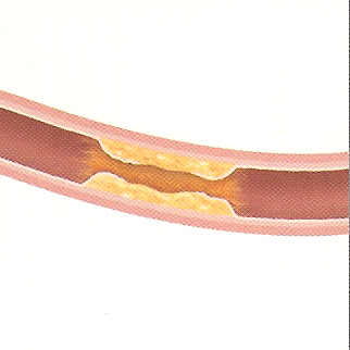 When
blood flow through an artery in the lower body becomes restricted, the blood
flow to lower extremities may be reduced or blocked. Insufficient blood
supply to legs or feet may cause discomfort, damage tissue, or even cause
gangrene.
When
blood flow through an artery in the lower body becomes restricted, the blood
flow to lower extremities may be reduced or blocked. Insufficient blood
supply to legs or feet may cause discomfort, damage tissue, or even cause
gangrene.
No one should ignore pain in their legs as it is
usually the first symptom of PAD. If walking even short distances is
uncomfortable, one should seek medical assistance.
Common exams used to determine the presence of
PAD are ABI (ankle brachial index) and arterial duplex. ABI is the most
common, the quickest and the least expensive exam to test for PAD. While
arterial duplex involves the use of an ultrasound machine, an ABI test is
performed through the use of blood pressure cuffs on arms and legs.
Treatment of PAD depends largely on how
constricted the artery has become. Treatment may range from lifestyle
changes (like proper diet or quitting smoking), exercise which improves
circulation, and medicine, to surgery if blood flow is severely hampered.
Imaging and physiologic tests help diagnose
arterial problems. They allow physicians to ascertain the severity of the
problem by viewing blood flow through the arteries. Lucid Diagnostic
Imaging specializes in providing doppler and duplex diagnostic exams to detect
blood flow and record images of arteries. Ultrasound procedures are
non-invasive and are painless. A technician moves a transducer over an
area of a patient's body to capture the images onto a machine. Those
images, along with a report, are then sent to an interpreting specialist who
views the images and report and performs an analysis of the study. The
interpreting physician makes his findings known to the ordering doctor after
he/she has completed the interpretation.
BACK